HKUMed innovates photocurrent-responsive coating: Cuts bone-to-implant integration time in half to just two weeks
12 December 2024
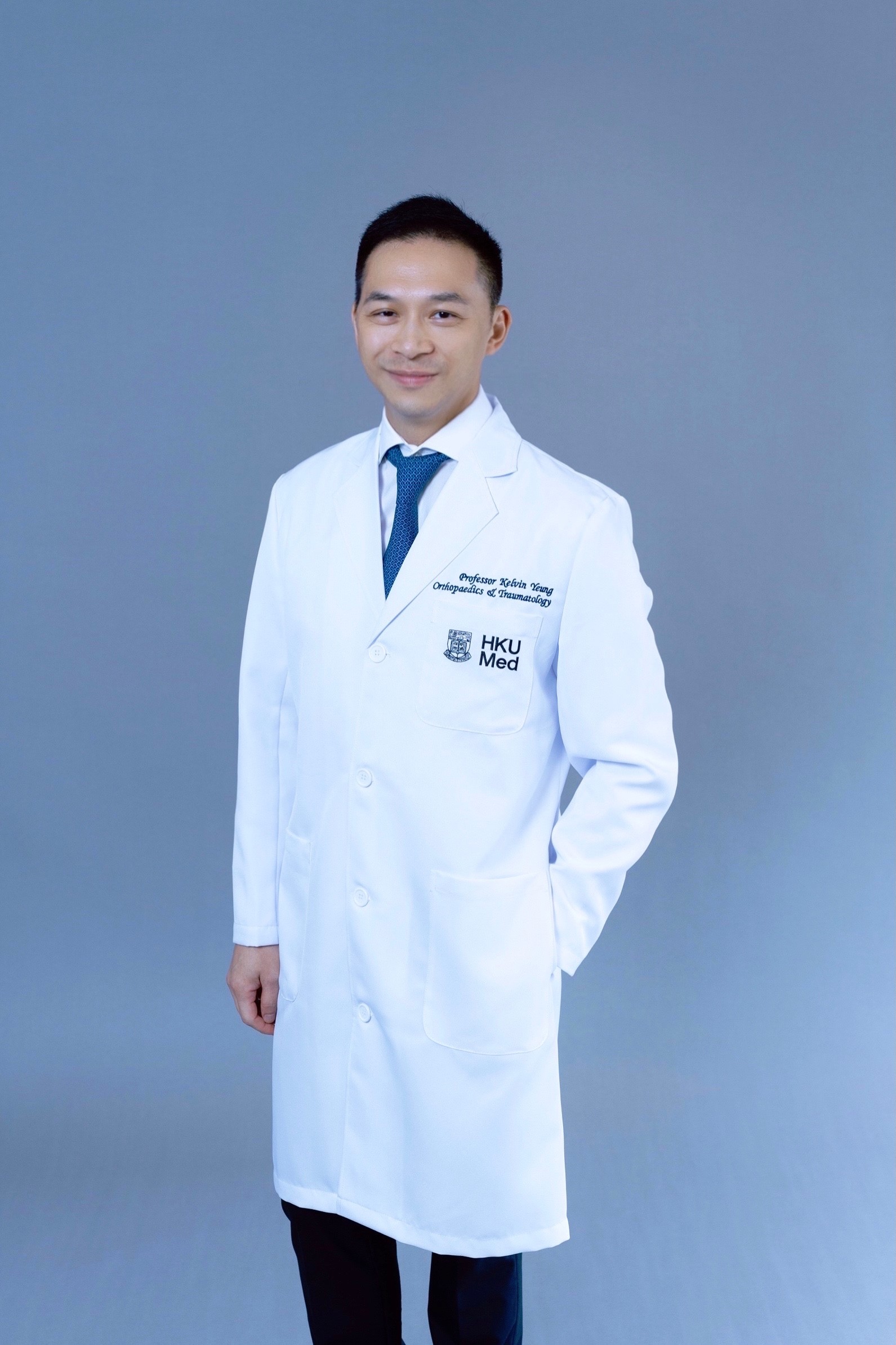
A research team led by Professor Kelvin Yeung Wai-kwok from the Department of Orthopaedics and Traumatology, School of Clinical Medicine, LKS Faculty of Medicine, the University of Hong Kong (HKUMed), has developed an innovative photocurrent-responsive implant surface to accelerate bone-to-implant integration after orthopaedic surgery. The cutting-edge coating has been shown to shorten the integration time to just two weeks, doubling the speed and expediting post-operative recovery as well as reducing the risk of rejection. The team is currently exploring the application of this technology in artificial joint replacement surgeries, including commonly performed knee replacement surgeries in Hong Kong.
Disorder in the osteoimmune microenvironment during the post-implantation phase can result in implant loosening, extending recovery time and increasing postoperative complications, ultimately resulting in implant failure. To address these challenges, the HKUMed team has developed a near-infrared (NIR) light-responsive implant surface that positively influences the macrophage response, effectively reducing acute inflammation during the crucial early post-implantation stage. This process involves the generation of a photocurrent in response to NIR light, which instigates increased calcium influx in macrophages, creating a more favourable osteoimmune microenvironment. This enhances the recruitment of mesenchymal stem cells (MSCs) and promotes bone formation, thereby speeding up the bone-to-implant integration process. The findings have been published in Advanced Functional Materials (link to publication).
Background
When an implant is introduced into the body, it triggers a complex response, known as foreign body reaction (FBR). This intricate process involves various cellular and molecular events that determine the effectiveness of bone-to-implant integration. Macrophages, which play a pivotal role in the FBR, are among the first immune cells to respond, initiating a chain reaction that is crucial for bone-to-implant integration. Upon insertion, these immune cells become activated and trigger an acute inflammatory response, releasing pro-inflammatory cytokines, like tumour necrosis factor-alpha, to facilitate the recruitment of mesenchymal stem cells (MSCs) and initiate the bone-regeneration process.
This inflammation process begins immediately upon implantation and peaks within a day or two. However, in a case in which the host immune system’s self-regulation is compromised due to a local pathological condition, the spike in inflammation may not be adjusted in time and may progress to chronic inflammation. This could result in a range of complications, including the formation of fibrous capsules, bone resorption, enzymatic degradation of implants, or a delay in integration, eventually leading to implant failure. Alarmingly, over 10% of implant failures are linked to implant loosening. Therefore, it is critical to restore a balanced environment between the bone and the implant, particularly after the initial inflammation phase, to prevent long-term inflammation and ensure successful implant integration.
Research findings and significance
Orthopaedic implants are typically coated with titanium dioxide (TiO2), which is non-toxic to bone cells and bacteria, but has limitations in its responsiveness to NIR. Known for its ability to penetrate biological tissues, NIR is widely used in antibacterial and cancer treatments. In this study, the research team used hydroxyapatite (HA), the primary component of bone and teeth, to develop an excitable surface that responds to a photocurrent. This novel coating generates photoelectric signals when exposed to NIR, which swiftly reduce acute inflammation and actively regulate macrophage differentiation, creating a beneficial immune environment tailored to the patient’s condition to foster bone-to-implant integration. This regulation promotes the recruitment of mesenchymal stem cells for bone formation, ultimately accelerating bone-to-implant integration, making the implants more secure.
In experiments using a tibial defect animal model, the team found that bone-to-implant integration was accelerated from 28 days to just 14 days, effectively doubling the speed. This marks the first study that uses a photocurrent to non-invasively regulate immune cells. This discovery is expected to advance the development of new biomaterials capable of remotely controlling the local immune environment.
‘Our team has successfully developed an engineered surface that non-invasively modulates macrophage differentiation according to the patient’s immune cycle and needs,’ said Professor Kelvin Yeung Wai-kwok, who led the research. ‘Animal experiments have proved that this method significantly accelerates bone-to-implant integration, resulting in a twofold increase in the fusion rate. We aim to expand the application of this engineered surface in orthopaedic surgeries in future research to enhance patient recovery. This discovery has a profound impact on the success rate of orthopaedic surgery and provides a new direction for addressing clinical challenges, like implant rejection.
About the research team
The research study was led by Professor Kelvin Yeung Wai-kwok, from the Department of Orthopaedics and Traumatology, School of Clinical Medicine, HKUMed. The first author, Dr Zhu Yizhou, is a postdoctoral fellow in the same department.
Acknowledgments
The research is jointly supported by the National Key Research and Development Programme of China, General Research Fund and Collaborative Research Fund from Research Grants Council (RGC), Innovation and Technology Fund, Health and Medical Research Fund, National Natural Science Foundation of China, Shenzhen Science and Technology Innovation Committee Projects, Guangdong Basic and Applied Basic Research Foundation, National Natural Science Foundation of China/ RGC Joint Research Scheme, and the National Science Fund for Distinguished Young Scholar.
Related media coverage
港大醫學院首創光電流感應塗層
骨科手術植入物融合只需兩周 速度提升一倍
2024年12月12日
香港大學李嘉誠醫學院(港大醫學院)臨床醫學學院矯形及創傷外科學系楊偉國教授率領團隊研發出一種新的光電流感應塗層,用於進行骨科手術,縮短植入物在體內與骨骼融合的時間至僅兩周,速度提升一倍並減少排斥。研究及實驗結果顯示,植入物與骨骼成功融合能加速患者術後康復。研究團隊計劃研究應用此技術於人工關節置換手術,例如本港甚為普遍的膝關節置換手術。
團隊發現,在現行的骨科手術中,當植入物植入患者體內後,骨免疫組織微環境的紊亂可能導致植入物鬆脫,有機會延長康復時間,引發術後併發症,某些情況下甚至會排斥植入物,導致手術失敗。因此,團隊研發了一種光電流感應塗層物料,可按需要調控巨噬細胞分化,減少植入物植入初期產生的急性炎症。該過程包括以近紅外光照射植入物表面塗層物料,產生光電流,引發巨噬細胞中鈣離子內流增加,從而創造一個更適合的骨免疫組織微環境,增強間充質幹細胞(Mesenchymal Stem Cells,MSCs)的募集達到成骨,有利骨骼與植入物融合。相關研究成果已於期刊《先進功能材料》發表(按此瀏覽期刊文章)。
研究背景
當患者身體植入金屬支架等異物時,會觸發異物反應(Foreign Body Reaction,FBR),過程涉及多種細胞及分子,影響骨骼與植入物的融合效果,當中名為巨噬細胞的免疫細胞是最先作出反應的細胞之一,並對後續骨骼與植入物融合的過程起決定性作用。植入物一經進入人體,這些免疫細胞會變得活躍,並隨即引發急性炎症反應,釋放免疫細胞激素,例如腫瘤壞死因子(Tumor Necrosis Factor-alpha),以促進間充質幹細胞募集並啟動骨骼再生。
炎症反應通常在植入手術後一至兩天最為嚴重,但每位患者的免疫周期各有不同,如因病理原因無法及時調節炎症,可能會發展為慢性炎症,導致纖維囊形成、骨再吸收、植入物酶降解,以及骨骼與植入物延遲融合等問題,最終導致身體排斥植入物。醫學文獻記載,超過10%的植入物排斥案例源於植入物鬆脫,因此在炎症初期主動調控病人骨骼和植入物之間的免疫組織微環境,能及時防止發展為長期炎症,對骨骼與植入物融合效果和穩定性至關重要。
研究結果及意義
現時廣泛用於骨科手術的鈦及其合金植入物表面具有一層自發形成的以二氧化鈦(TiO2)薄膜,雖然對骨細胞和細菌無毒,但TiO2的特性限制了其對近紅外線(Near Infrared,NIR)回應的性能;而近紅外線因為能穿透生物組織而廣泛應用於抗菌和癌症治療。在這項研究中,港大醫學院團隊使用羥基磷灰石(組成骨骼和牙齒的主要成分)為原材料,研發出一種能與光電流感應的植入物塗層,這個塗層經近紅外線照射時便會產生光電信號,及時減輕急性炎症,主動調控巨噬細胞分化行為,根據患者情況建立有利的免疫環境促進骨骼與植入物融合。這樣的調節能促進間充質幹細胞的招募達到成骨,最終加快骨骼與植入物的融合速度,令植入物更加鞏固。
團隊在研究脛骨缺陷動物模型中發現,這種新塗層顯著加速骨骼與植入物融合,由28天縮短至14天,足足一倍之多。這是首次利用光電流以非侵入性方式調節免疫細胞的研究,這項發現有望推動新型生物材料的開發,使其能夠遠程控制局部的免疫環境。
楊教授說:「團隊研發出的人工塗層,成功以非侵入性方式,按患者的免疫周期和需要調控巨噬細胞的分化行為,並在動物實驗中證實這種方法能顯著加速骨骼與植入物的融合達一倍。我們期望在未來研究中,進一步開拓此人工塗層在骨科手術上的廣泛應用,從而提升患者的術後康復效果。這不僅對骨科手術的成功率有著深遠影響,也為解決植入物排斥等臨床問題提供嶄新的方向。」
研究團隊
這項研究由港大醫學院臨床醫學學院矯形及創傷外科學系教授楊偉國教授領導。第一作者為同學系的博士後研究生祝亦周。
鳴謝
此項研究獲得多項資助,包括國家重點研發計劃、研究資助局的「優配研究金」和「協作研究金」、創新及科技基金、醫療衞生研究基金、中國國家自然科學基金、深圳市科技創新委員會專案、廣東省基礎與應用基礎研究基金、國家自然科學基金委員會及研究資助局聯合科研資助基金、國家傑出青年科學基金項目。

